Small is beautiful, as winners of this Nikon International Small World competition prove.
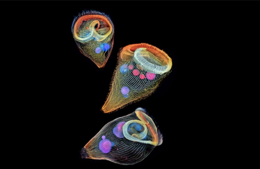
First prize in the 2019 Photomicrography Competition went to a picture of a fluorescent turtle embryo taken by Teri Zgoda and Teresa Kugler from New York. The pair used a technique called image stitching to make a composite from hundreds of images.
“We are inspired by the beautiful images we see through the microscope,” Zgoda said. “It’s humbling and deeply fulfilling to be able to share that science with other people.”
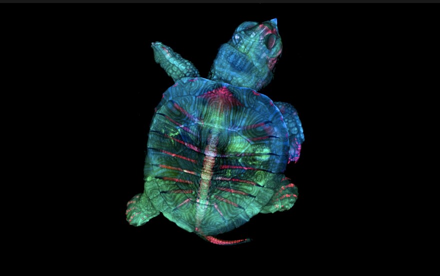
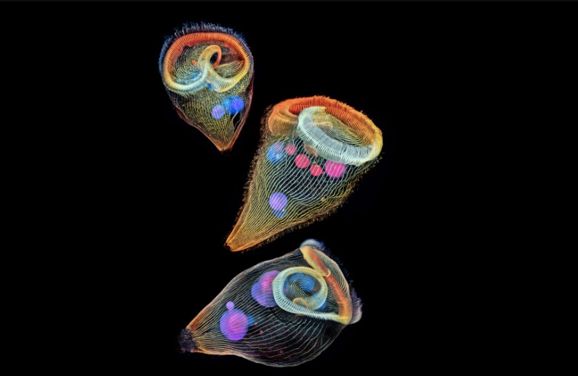
Second prize went to Dr Igor Siwanowicz for his “depth-colour coded projections of three stentors (single-cell freshwater protozoans)”. Stentors, also known as trumpet animalcules, are filter-feeding ciliates – creatures with hair-like appendages called cilia. Although they are undoubtedly tiny, at around two millimetres in length, these stentors are some of the largest single-cell organisms.
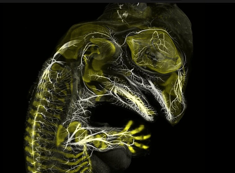
Third place went to Daniel Smith Paredes and Dr Bhart-Anjan S Bhullar. Their photograph of an alligator embryo shows the creature’s skeleton and nerves forming.
If you’ve ever had the feeling that someone is watching you, the sixth-placed photo might unnerve you. It shows a small white hair spider in stunning close-up.

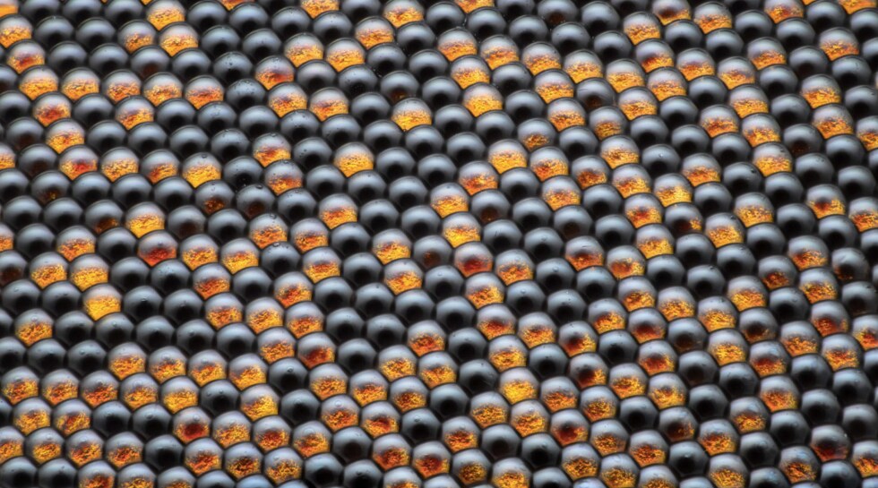
Or this extreme close-up of the eye of a housefly.
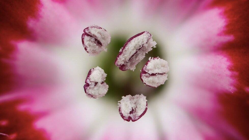
This photo might look like something on a cosmic scale, but is actually the stamen of a flower.
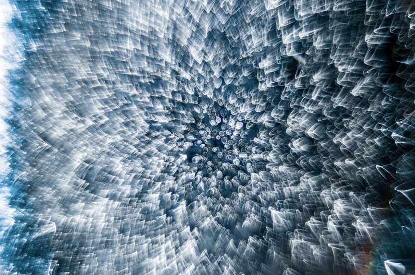
If you like the magic of the microscopic, you’ll probably appreciate this photo of a frozen droplet of water.
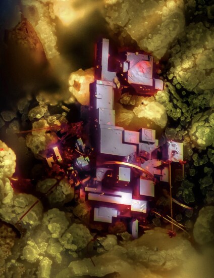
Or perhaps this architectural-looking close-up of some cuprite — copper oxide.
The top 20 are exhibited at museums and science centers across North America.